How to Tell Annelids, Arthropods, Molluscs and Echinoderms Apart for NEET
A specimen with repeated body units might be an annelid or an arthropod; a soft body might suggest a mollusc until an internal skeleton and radial arrangement point to an echinoderm. The reliable way to separate these four phyla is to use a fixed sequence: inspect segmentation, ask whether any appendages are jointed, determine the body symmetry, and then confirm the identification with a signature structure. This works better than relying on habitat, shell, or overall shape, because members of the same phylum can look strikingly different.[1][2][3]
A four-character sequence turns resemblance into identification
Begin with segmentation, meaning the repetition of body units. Annelids have true metameric segmentation: internal and external features repeat through successive body segments.[1][4] Arthropods are also segmented, but adjacent segments may fuse into functional regions called tagmata, such as a head, thorax and abdomen or a cephalothorax and abdomen.[2] Molluscs instead share a body plan centred on a muscular foot, visceral mass and mantle, while adult echinoderms are organised around a central axis rather than as a chain of repeated segments.[1][3]
Next examine appendages. Jointed appendages are a principal feature of arthropods and may be specialised for sensing, feeding, walking, jumping, digging or swimming.[2] Annelid parapodia, when present, are paired but unjointed, so an appendage is not automatically an arthropod leg.[4] Molluscan arms and tentacles can be modifications of the foot, while echinoderm tube feet belong to a water vascular system; neither should be confused with arthropod jointed appendages.[1][3]
Then use symmetry. Annelids and the basic molluscan body plan are bilateral, with left and right sides arranged around one main plane.[1] Adult echinoderms usually show pentaradial symmetry, with body parts commonly arranged in multiples of five around a central axis, although their larvae are bilateral.[3] Arthropods may differ greatly in outline, but their segments and paired appendages remain the more decisive characters supplied by the sources.[2]

Four visible checks narrow the specimen to one phylum
Think of this sequence as a lock with several tumblers. That is an analogy, not a biological finding: a single matching feature suggests an answer, but the complete combination makes the identification dependable.
Repeated rings plus chaetae identify an annelid
Annelids are bilaterally symmetrical, worm-like animals whose true segmentation repeats internal and external features from one body segment to the next.[1][4] Membranous partitions divide the coelomic cavity into a series of compartments, and several organ systems also follow the segmental arrangement.[1] Their body can be divided broadly into a head, segmented body and tail region.[1][4]
For an exam identification, the most useful signature structure is the chaeta, also called a seta: a chitinous bristle anchored in the body wall.[1][4] Most annelid groups bear chaetae in their segments.[4] In polychaetes, clusters of these bristles occur on paired parapodia, which are fleshy, unjointed appendages used in locomotion and breathing.[1][4] Earthworms have fewer, reduced chaetae and also possess a clitellum, the conspicuous ring involved in reproduction.[1][4]
The important trap is the leech. Leeches are annelids even though they lack chaetae and instead possess anterior and posterior suckers.[1][4] Their visible body-wall rings may also fail to match the internal segmentation of the body cavity exactly.[1][4] Therefore, chaetae are a strong positive clue, but their absence does not by itself exclude Annelida.
Illustrative example: imagine a bilaterally symmetrical worm with a chain of ring-like segments and tiny bristles, but no articulated legs. The repeated compartments establish true segmentation, the lack of jointed appendages separates it from an arthropod, and the chaetae confirm the annelid pattern.[1][2][4]

True body segments and chaetae form the main annelid pattern
Jointed appendages make an arthropod unmistakable
Arthropods combine body segmentation with jointed appendages and a chitinous exoskeleton.[2] Of these characters, jointed appendages provide the quickest separation from the other three phyla.[2] Even when the body segments have fused into tagmata, the appendages retain the arthropod pattern and may be modified into antennae, mouthparts, walking legs or other specialised structures.[2]
The signature structure is the chitinous exoskeleton, a tough external covering rather than an internal skeleton.[2] Because this covering cannot simply expand continuously, arthropods grow through molting and then ecdysis, the actual shedding of the old exoskeleton.[2] This contrasts directly with the echinoderm endoskeleton, whose calcareous plates lie beneath the epidermis.[3]
Different arthropods arrange their segments and legs differently. Insects have a head, thorax and abdomen, with three pairs of jointed appendages on the thorax.[2] Centipedes bear one pair of legs per segment, whereas millipedes bear two pairs on each diplosegment formed by the fusion of adjacent segment pairs.[2] Many crustaceans have a cephalothorax and abdomen, while their appendages commonly divide into two branches.[2] These variations alter the layout, not the underlying combination of segmentation, articulation and external skeleton.[2]
Illustrative example: a specimen has visible body regions rather than a simple series of identical rings, and its legs bend at distinct joints. The jointed legs identify the arthropod pattern; a rigid chitinous covering supplies confirmation.[2] A bristled parapodium would point elsewhere because annelid parapodia are unjointed.[4]

Jointed appendages separate arthropods from superficially segmented annelids
A mantle-based body plan reveals a mollusc
Molluscs vary from snails and clams to squids and octopuses, yet their shared plan includes a ventral muscular foot, a visceral mass containing most internal organs, and a dorsal mantle over the visceral mass.[1][4] The mantle creates the mantle cavity, which commonly houses respiratory structures and other openings.[1][4] The foot may serve in locomotion or anchorage, become a crawling surface, or be modified into the arms, tentacles and funnel of a cephalopod.[1][4]
The mantle is the safest signature structure because neither a shell nor a radula is universal. In most molluscs, the mantle secretes a calcium-carbonate shell, but the shell is reduced in squids and cuttlefish and absent in octopuses and slugs.[1][4] Most molluscs possess a rasp-like feeding organ called a radula, yet bivalves lack it.[1][4] A rule based only on “shell present” or “radula present” would therefore produce false exclusions.
Segmentation is also a poor route to Mollusca. The useful recognition pattern is an unsegmented-looking body organised around the foot, visceral mass and mantle rather than a serial chain of repeated compartments.[1][4] Molluscs do not have the jointed appendages that characterise arthropods.[1][2] Their basic plan is bilateral, although gastropod bodies become asymmetrical and commonly carry a coiled shell.[1]
Illustrative example: a soft-bodied animal has no repeated rings and no jointed legs, but a broad ventral surface lies beneath a mass of organs covered by a tissue fold. The ventral structure fits the muscular foot, and the covering fold fits the mantle, so Mollusca is the supported identification.[1][4]
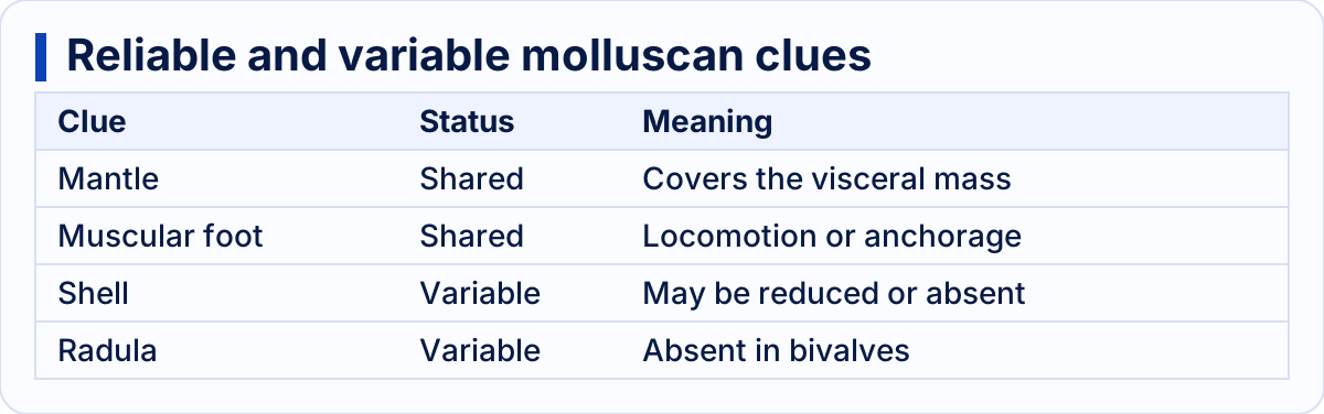
The mantle confirms molluscs even when shell and radula are absent
Five-part adult symmetry and tube feet point to an echinoderm
Adult echinoderms generally exhibit pentaradial symmetry: their parts are arranged around a central axis, usually in multiples of five.[3] Sea stars display the pattern through their arms, while sea urchins and sand dollars lack arms but retain five rows of tube feet passing through rows of pores.[3] Echinoderm larvae are bilaterally symmetrical, so the radial condition belongs to the adult stage rather than the whole life cycle.[3]
Their signature structure is the water vascular system, a fluid-filled system derived from the coelom.[3] It includes a central ring canal, radial canals and connected tube feet.[3] Changes in water volume extend or retract the feet, which can assist in movement, attachment, feeding and sensation.[3] Seawater enters through the madreporite and reaches the ring canal through the stone canal.[3]
A second confirming feature is an endoskeleton of calcareous ossicles—small plates located beneath the epidermis.[3] This placement distinguishes it from the external chitinous skeleton of arthropods.[2][3] The ossicles may be fused, separately embedded in connective tissue or reduced to tiny spicules, as in sea cucumbers.[3]
The chief symmetry trap is the adult sea cucumber. Its elongated body gives it functional bilateral symmetry, and its tube feet may be reduced or absent except on the side against the substrate.[3] In such a case, the water vascular system and internal ossicles are more dependable than the external outline.
Illustrative example: a marine animal has five radiating regions, rows of tube feet and no jointed legs. The adult pentaradial arrangement suggests Echinodermata, while the tube feet connected to a water vascular system confirm it.[3]

Adult radial symmetry is confirmed by the echinoderm water vascular system
The decisive comparison is a combination, not one isolated clue
A segmented body alone cannot separate annelids from arthropods because both phyla are segmented.[1][2] The discriminator is articulation: arthropods possess jointed appendages, whereas annelid parapodia are unjointed and commonly bear chaetae.[2][4] Likewise, a hard covering alone is ambiguous: molluscs may have a mantle-secreted shell, arthropods have a chitinous exoskeleton, and echinoderms possess calcareous ossicles beneath the epidermis.[1][2][3]
The following is a study rule based on the sourced characters, not an additional biological finding: identify the broad body organisation first and use the signature structure as confirmation. “Segmented worm” should lead to chaetae; “segmented animal with articulated legs” to the exoskeleton; “foot–visceral mass–mantle plan” to the mantle; and “five-part adult organisation” to the water vascular system.
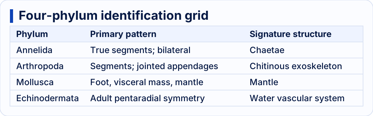
Each phylum has a distinctive four-character combination
Key takeaways
- Annelids have true repeated segments, bilateral symmetry and usually chaetae; any parapodia are unjointed.[1][4]
- Arthropods combine segmentation with jointed appendages and a chitinous exoskeleton that is shed during growth.[2]
- Molluscs are recognised by the muscular foot, visceral mass and mantle; shells and radulae are not universal.[1][4]
- Adult echinoderms usually have pentaradial symmetry, an internal skeleton of ossicles and a water vascular system connected to tube feet.[3]
- Exceptions must refine the rule rather than erase it: leeches lack chaetae, bivalves lack a radula, several molluscs lack an external shell, and adult sea cucumbers show functional bilateral symmetry.[1][3][4]
This comparison settles how to distinguish the four phyla using the requested anatomical characters. It does not classify every subgroup within them; unusual specimens may require several internal characters because a conspicuous external clue can be reduced, modified or absent.[1][2][3][4]
Sources
- https://openstax.org/books/biology-2e/pages/28-4-superphylum-lophotrochozoa-mollusks-and-annelids (retrieved 2026-09-30)
- https://openstax.org/books/biology-2e/pages/28-6-superphylum-ecdysozoa-arthropods (retrieved 2026-09-30)
- https://openstax.org/books/biology-2e/pages/28-7-superphylum-deuterostomia (retrieved 2026-09-30)
- https://openstax.org/books/concepts-biology/pages/15-4-mollusks-and-annelids (retrieved 2026-09-30)